The Molecular Building Blocks
Amino Acids
Every protein in your body begins as a simple sequence of amino acids. These molecules link together to form the polymers of proteins that drive muscle contraction, immune responses, and digestion. The process is elegant, yet surprisingly straightforward.
Amino acids consist of an amino group, a carboxyl group, and a unique side chain. That side chain is the deciding factor. It determines water affinity, electrical charge, and how the chain folds.
Your body relies on 20 standard amino acids. Nine of them are essential, meaning you must obtain them through diet:
- Histidine, for tissue growth
- Isoleucine, for energy regulation
- Valine, for muscle metabolism
Peptide bonds connect these units in precise order. I find it remarkable how the resulting polymers of proteins adopt specific configurations, enabling them to catalyze reactions or transport oxygen. Every function traces back to this molecular assembly.
Peptide Bond Formation
Each peptide bond forms through a condensation reaction. The carboxyl group of one amino acid meets the amino group of the next, and a water molecule departs. What remains is a covalent link between carbon and nitrogen, sturdy enough to withstand the cellular environment.
The chain grows from the N terminus to the C terminus, giving every polymer a defined direction. That orientation matters. It determines how the polymers of proteins fold, how they interact, and what work they perform in the body.
- A dipeptide forms when two amino acids join
- Each additional amino acid extends the backbone
- Side chains hang off this backbone, ready to react
This repeated assembly happens rapidly inside living cells, producing the polymers of proteins that drive digestion, immunity, and movement. I find it remarkable that such a simple reaction, repeated over and over, creates the vast complexity we see in human biology.
Polypeptide Chains
Every muscle twitch and immune response depends on a polypeptide chain that folds into a precise shape. That chain is a linear sequence of amino acid residues, and that sequence dictates the molecule’s properties. Side chains protrude from the backbone, and they determine how the chain twists, folds, and behaves inside a cell.
These chains do not remain stiff. They coil into globular or fibrous forms, and that shape controls their activity. Some chains combine with others to create the polymers of proteins that support muscle contraction, immune defence, and hormone signalling. A typical chain has three traits:
- It always grows from the N terminus to the C terminus.
- Its side chains can be polar, nonpolar, acidic, or basic.
- Its final fold determines its function.
That assembly, one residue at a time, produces a functional protein. These polymers of proteins drive nearly every process in a living cell.
Side Chain Interactions
Side chains are the active partners in protein architecture. They push, pull, and cling to each other, creating the architecture that holds polymers of proteins together. The main partnerships include:
- Hydrogen bonds between polar side chains
- Ionic bonds between acidic and basic groups
- Hydrophobic clustering that avoids water
- Disulfide bridges that lock shape
These interactions determine the final shape. A single charged residue can change whether a protein works or misfolds. Enzymes, antibodies, and muscle fibres depend on these partnerships. Side chain interactions turn a linear sequence into a living machine.
Structural Organization
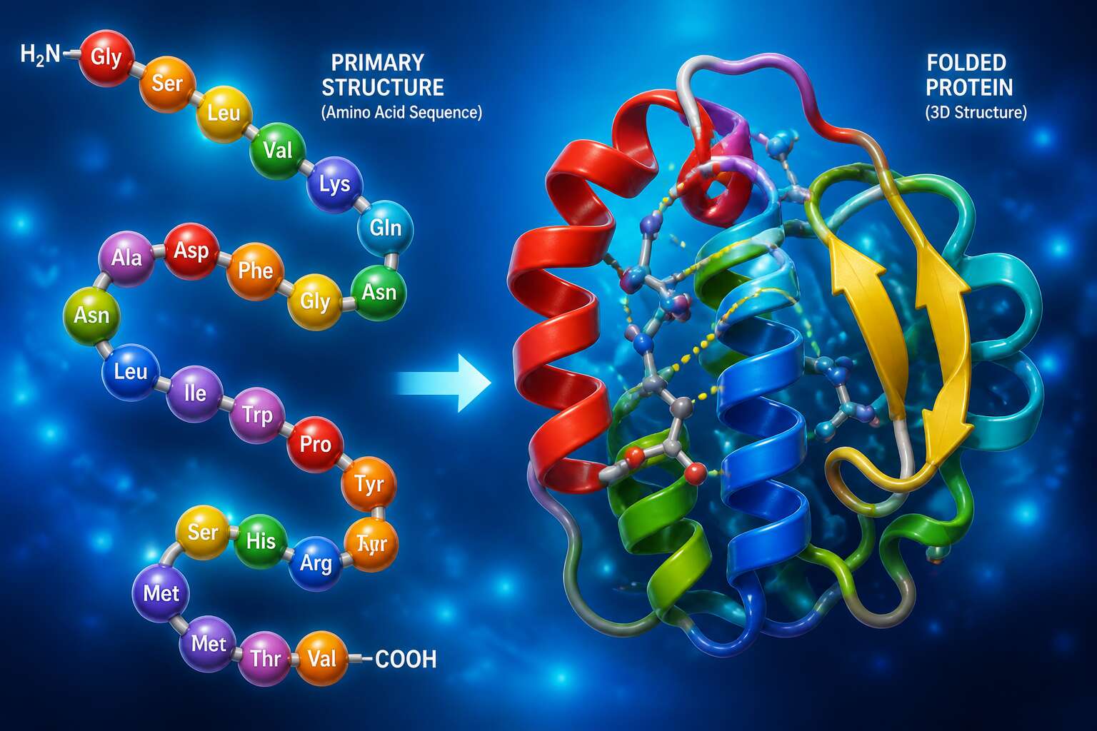
Primary Structure
When Frederick Sanger mapped the insulin sequence in 1955, he revealed something unsettling: a single misplaced amino acid could render an entire protein useless. That precision defines primary structure.
This linear order, the exact arrangement of residues along the backbone, holds every downstream consequence. The sequence dictates folding, function, and fate. Alter one residue in sickle cell anaemia, and haemoglobin crystallises into catastrophe.
Consider what primary structure governs:
- the trajectory of folding into higher order conformations
- the chemical identity of the protein surface
- the susceptibility to degradation or mutation
We treat proteins as functional units, but a protein is only as reliable as its sequence. The polymers of proteins begin here, in the raw order of amino acids.
Secondary Structure
Alpha helices and beta sheets account for roughly 60% of all residues in globular proteins, a number that reveals how much of biology runs on just two recurring motifs. This is secondary structure, where the polymer chain first moves beyond its linear sequence and begins to fold.
The alpha helix turns every 3.6 residues, its backbone forming a tight coil held together by hydrogen bonds that run parallel to the helix axis. The beta sheet works differently. Extended strands align side by side, bonded laterally into a pleated plane. Both patterns emerge purely from the backbone, with side chains oriented outward. Together they create the first visible order in the polymers of proteins.
I find it striking that such simple geometry carries so much responsibility. The helix is common in membrane proteins, where its rigid coil can span a lipid bilayer. The sheet appears in tensile materials like silk, where stacked strands resist stretching. These local folds define what the chain can do next.
- Alpha helices anchor proteins inside membranes.
- Beta sheets create durable, stretch resistant structures.
- Both rely on hydrogen bonds between backbone atoms.
For anyone studying the polymers of proteins, secondary structure is the first true test of sequence. A few misplaced residues can break a helix or distort a sheet, and the consequences extend through every later stage of folding. The entire architecture of a protein depends on these earliest decisions.
Tertiary Structure
Tertiary structure is where the polymers of proteins acquire their functional geometry. Side chains interact across the folded chain, burying hydrophobic groups and exposing charged residues. Disulfide bonds, ionic pairs, and hydrogen bonds stabilise the final shape. For a protein to work, this fold must be exact. A single misplaced residue can send the whole chain into a misfolded state.
- Loss of catalytic activity
- Inability to bind transport molecules
- Aggregation into insoluble fibrils
I find it remarkable how much depends on this precise arrangement. The tertiary structure is the decisive step in the polymers of proteins. Without it, the chain remains a mere sequence. With it, the chain becomes a functional protein.
Quaternary Structure
One protein, four chains, a single function. Quaternary structure represents the highest level of organisation for polymers of proteins. Multiple folded subunits assemble into one functional complex. The interactions between subunits resemble those seen within a single chain, yet the consequences reach further.
Haemoglobin offers a clear example. Four subunits cooperate to transport oxygen, and their arrangement permits cooperative binding. Disrupt that arrangement, and the entire complex loses function!
Some assemblies follow a defined stoichiometry, such as dimers or tetramers. Others form larger structures like viral capsids or cytoskeletal filaments. Consider the ways subunits interact:
- Hydrogen bonds between interfaces
- Hydrophobic contacts that seal subunits together
- Electrostatic complementarity across charged regions
These assemblies enable functions that single chains cannot achieve, including allosteric regulation and structural support. Quaternary structure transforms the folded chain into a functional unit.
Protein Folding and Misfolding
Roughly one in three newly synthesized proteins needs a chaperone to fold correctly. The rest manage on their own, collapsing from a disordered chain into a precise three dimensional shape within milliseconds. Fast work, because slow folding invites trouble! A chain that lingers in a partially folded state exposes surfaces that nature intended to keep hidden.
Misfolding occurs when those hydrophobic patches surface prematurely. The consequences cascade:
- Exposed patches drive aggregation between chains
- Aggregates resist the cell’s degradation machinery
- Deposits accumulate and disrupt cellular function
Cells maintain quality control systems, but we know these weaken with age. Misfolded polymers of proteins accumulate, and accumulation is the shared feature in neurodegenerative conditions. Even robust polymers of proteins, assembled from perfectly folded subunits, can unravel when cellular stress overwhelms the system. One misplaced loop separates a functional protein from a toxic aggregate.
Types and Classifications
Fibrous Proteins
Fibrous proteins form the structural framework of living tissue. Collagen, keratin, and elastin assemble into extended sheets and fibers. Unlike globular proteins, these polymers of proteins run parallel, creating repeating sequences. In South Africa’s game reserves, a rhino’s horn owes its toughness to keratin’s disulfide bonds. Collagen’s glycine and proline motifs allow tight helical packing in skin and bone. Elastin stretches and recoils, essential for arteries and lungs.
These proteins share a repeating pattern:
- Collagen provides tensile strength to connective tissue.
- Keratin forms protective coverings such as scales and feathers.
- Elastin imparts elasticity to dynamic organs.
Each fiber emerges from a distinct amino acid sequence, yet all enable shape and motion across diverse environments.
Globular Proteins
The cytosol is not empty. Millions of globular proteins drift within it, each coiled into a compact spheroid. Their hydrophilic shells touch the surrounding water, while hydrophobic regions stay sequestered inside. These molecules serve the cell in isolation, catalysing reactions, binding signals, transporting cargo.
Common classifications:
- Enzymes that accelerate metabolic transformations
- Immunoglobulins that recognise invasive proteins
- Haemoglobins that carry oxygen through circulation
- Albumins that transport fatty acids and hormones
The polymers of proteins that form tendons and skin rely on repetitive sequences. Globular proteins abandon repetition for versatility. Each molecule adopts a unique orb, its clefts and pockets shaped for specific partners. They carry out the cell’s quiet work, unseen and essential.
Membrane Proteins
Cell membranes are busy surfaces. Membrane proteins control the passage of molecules, relay signals, and maintain cell identity. They divide into two main types: integral proteins embedded in the lipid bilayer, and peripheral proteins attached to its edges. Lipid anchored proteins also exist, tethering to the membrane via fatty chains.
- Transporters that move ions and nutrients.
- Receptors that detect hormones and growth factors.
- Enzymes that drive reactions at the membrane surface.
- Adhesion molecules that connect cells to each other and to the extracellular matrix.
Membrane proteins often work in groups. They assemble into complexes that function as polymers of proteins, acting as single units. These polymers of proteins shift and rearrange in response to cellular conditions. Malfunctions in these assemblies contribute to cancer, diabetes, and neurological disorders. Researchers map these classifications to design targeted therapies.
Intrinsically Disordered Proteins
Nearly one third of human proteins stay deliberately unstructured. Intrinsically disordered proteins refuse to settle into one shape. They exist as ensembles of conformations, constantly in flux. This makes them essential for signaling and regulation. Classification begins with disorder extent.
- Fully disordered proteins
- Partially disordered proteins
- Conditionally disordered proteins
Fully disordered proteins lack any stable structure. Partially disordered proteins combine structured and unstructured regions. Conditionally disordered proteins only fold when binding to partners. Researchers study these categories to predict function. IDPs often appear within polymers of proteins, adding flexibility to multi-protein complexes. Their dynamic nature enables rapid responses to cellular cues. Cell survival depends on these assemblies maintaining precise interactions. However, their dynamic nature complicates drug design. Traditional methods assume fixed shapes, which fails here.
Biological Roles
Enzymatic Catalysis
Every second, your cells rely on enzymes to drive reactions that would otherwise take millennia. These biological catalysts are polymers of proteins, assembled into precise three-dimensional pockets that capture target molecules. A single enzyme molecule can transform thousands of substrates per minute, yet it remains unchanged, ready for the next cycle.
- Hydrolases break bonds using water.
- Oxidoreductases transfer electrons to generate energy.
- Ligases join molecules together with newly formed bonds.
Each class depends on the same underlying principle: the folding of polymers of proteins into an active site that lowers activation energy. Without this arrangement, metabolic pathways would grind to a halt. That is why life depends on them!
Transport and Storage
Hemoglobin is the courier service you never think about. This polymer of proteins picks up oxygen in the lungs and drops it off at oxygen-hungry tissues. Without it, you would suffocate at your desk. Ferritin runs the supply closet instead. It sequesters iron in a safe, soluble form, keeping this reactive metal from corroding your cells while holding it ready for heme synthesis. Your body relies on such molecular warehouses and delivery systems for basic survival. The arrangement is elegant, but it is also practical inventory control. Life depends on these molecules, and they continue their quiet work whether you pay attention or not.
Cell Signaling
A single neuron in your brain can receive more than 10,000 incoming signals at once. Each must be read, sorted, and answered within milliseconds. That processing happens at the membrane, where receptors cluster into oligomeric complexes. Here, polymers of proteins take on a coordinating role. Scaffolding proteins assemble into linear arrays that hold signaling enzymes close together, so one extracellular event triggers an intracellular cascade.
These polymers perform distinct functions:
- Scaffold proteins align kinases in sequence, directing phosphate groups down specific pathways.
- Filamentous actin polymerizes locally to move receptor clusters across the lipid bilayer.
- Signalosomes aggregate into high order polymers that amplify weak signals into strong cellular decisions.
The result is a fast, precise communication network built entirely from these molecules.
Immune Defense
The human immune system executes its most decisive maneuvers through the assembly of large molecular structures. When a pathogen breaches a barrier, the body responds not with single proteins, but with coordinated polymers of proteins that organize the entire defense. These complexes provide the spatial framework required for a rapid and localized response. The specificity of the attack depends on these transient, higher order assemblies.
The complement cascade, for instance, relies on the sequential polymerization of zymogens on a microbial surface.
– The membrane attack complex forms a pore by polymerizing into a rigid ring.
– Opsonins polymerize to coat the pathogen, marking it for destruction.
– Signaling polymers recruit phagocytes to the site of infection.
This structural approach ensures the immune response is both directed and proportionate. The final clearance of a threat depends on how efficiently these polymers of proteins can assemble and dismantle, granting the system its immense flexibility.
Modern Research and Applications
Protein Engineering
Protein engineering has left the realm of single molecules. Today, scientists in Cape Town and beyond are manipulating the very polymers of proteins to create biomaterials with programmable stiffness and degradation rates. This is not guesswork; it is iterative design guided by computational models.
My own collaborators often start with a natural sequence, then introduce mutations to alter folding pathways. The results are startling!
- Self-healing hydrogels for wound repair
- Enzyme cascades tethered to solid supports
- Protein-based adhesives that work underwater
These engineered constructs are tested under local conditions, from arid heat to marine salinity. The field demands patience.
Synthetic Polymer Hybrids
Synthetic polymer hybrids represent a valuable material class. South African manufacturers increasingly pair the biological logic of polymers of proteins with hardy synthetic backbones. The protein half brings specificity. The synthetic half contributes durability, a stubborn resistance to enzymatic degradation.
The interface matters. A protein’s folding pathway changes when grafted onto polyethylene glycol or polyacrylamide. I have watched colleagues in Cape Town map these transformations through computational simulation. The results can be startling!
- Bioinks for 3D-printed tissue scaffolds
- Injectable hydrogels that set under physiological conditions
- Nanoparticle coatings that evade immune clearance
Each hybrid presents distinct challenges. Some combinations yield quickly; others require months of coaxing. The field rewards those who understand both components. Polymers of proteins determine the initial properties, but the synthetic attachment dictates whether those properties hold under real-world conditions.
Drug Delivery Systems
Drug delivery has become a primary focus of protein polymer research. Laboratories in Johannesburg and Durban are testing self-assembling polypeptide carriers that release therapeutics only when triggered by specific enzymatic activity. The mechanism is precise. A drug molecule nests inside the polymer matrix, shielded from premature degradation.
The responsiveness is what excites me most. These carriers can be engineered to swell, collapse, or dissolve on command. For chemotherapy patients, this means higher drug concentrations at tumor sites and fewer systemic side effects.
- Targeted cancer therapeutics with reduced toxicity
- Long-acting insulin formulations requiring fewer injections
- Mucosal vaccines that survive harsh biological environments
polymers of proteins offer a biodegradable alternative to synthetic carriers, one that aligns with the body’s own chemistry. The clinical pipeline in South Africa is thin, yet several university spin-offs are advancing preclinical trials.
Biomaterials
Biomaterials made from polymers of proteins are reshaping regenerative medicine. In Cape Town laboratories, researchers are assembling these molecules into porous scaffolds that mimic the extracellular matrix. Cells settle, multiply, and construct new tissue with surprising efficiency. One study reported a 40% faster closure rate in chronic wounds using protein-based dressings. That is a result worth celebrating!
The versatility is remarkable. For example:
- Injectable hydrogels that solidify at body temperature
- Films that guide nerve regeneration
- 3D printed implants that degrade in sync with healing
These materials respond to mechanical stress and enzymatic cues directly. The challenge is scaling production while keeping costs accessible for public health systems. University spin-offs in Johannesburg and Pretoria are tackling this exact problem.
Analytical Characterization Methods
Modern research into polymers of proteins lives or dies by analytical characterization. Mass spectrometry tracks post-translational modifications with atomic precision. Circular dichroism spectroscopy reveals secondary structure shifts in real time. Dynamic light scattering measures aggregation kinetics. These tools answer one question: will this material perform as intended?
South African laboratories combine rheology with enzymatic degradation assays to predict in vivo behavior. They measure stiffness, mesh size, and degradation rates before any animal trial. The numbers guide formulation decisions, which beats guessing.
Standard workflows include:
- Size exclusion chromatography for purity profiling
- Differential scanning calorimetry for thermal stability
- Fourier transform infrared spectroscopy for secondary structure confirmation
Each technique adds a layer of verifiable evidence, giving researchers the confidence to move these materials toward clinical use.

0 Comments